World of Ophthalmology Dr. Victor Zamyatin's Personal Web Site
| Ophthalmology - Home Page | Anatomy | Physiology | Diseases | Diagnostic Tests | Surgical Procedures |
| Medications | Contact Lenses & Eyeglasses | Journals | Images | Links | FAQs | News |
| Congresses, Events | Trials | Eye Banks | Equipment | Eye & Vision Companies | Contact |
|
Encyclopaedia of Ophthalmology - Greatest Links' Collection |
|
Look 'World of Ophthalmology' on http://www.wophth.com ! |
|
Advertising
Banners appear in the left column |
|
► Anatomy |
|
► Diseases |
|
► Surgery |
 |
| We subscribe to the HONcode principles . Verify here |
|
'World of Ophthalmology' depends on donations and sponsorship. Sponsors will be offered the opportunity to
advertize/display their brand |
||||||||||||||
| Catalogue of Ophthalmic Images | ||||||||||||||
|
|
||||||||||||||
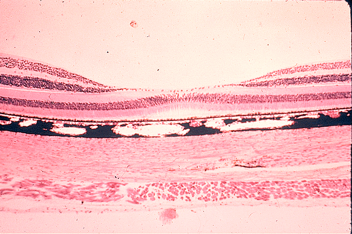
| Sclera | ||||||||||||||
|
2 Eyes - Anatomy / ► Sclera |
||||||||||||||
| Dr. Charles J. Pavlin's Ultrasound Biomicroscopy Web Site | ||||||||||||||
| ► |
||||||||||||||
| FotoWeb - Ophthalmic Images | ||||||||||||||
| ► Sagittal cut of the eyeball. Detail of sclera and optic nerve | ||||||||||||||
| Macula.org | ||||||||||||||
| ► Sclera | ||||||||||||||
| St Luke's Cataract & Laser Institute - Anatomy Focus | ||||||||||||||
| ► Sclera | ||||||||||||||
| Suny Downstate Medical Center | ||||||||||||||
| ► Sclera | ||||||||||||||
| U. of Delaware, Department of Biological Sciences, Mammalian Histology (B408) | ||||||||||||||
| ► Fovea Centralis, Choroid, & Sclera | ||||||||||||||
| U. of Texas Medical Branch, Cell Biology Graduate Program, Microanatomy Web Atlas | ||||||||||||||
| ► Retina, Choroid, and Sclera | ||||||||||||||
| Vision CyberMedia Group, Development of the Eye Site | ||||||||||||||
| ► Figure 12.
Photomicrograph of a sagittal section of the developing eye at about 50 days.
► Figure 5. Successive sagittal sections of developing eye at: A, 5 weeks B, 6 weeks C, 20 weeks D, Newborn ► Figure 11. Sagittal section through the developing eye at 15 weeks. |
||||||||||||||
| Xalatan Image Library | ||||||||||||||
| ► The Sclera | ||||||||||||||
| ► The Sclera | ||||||||||||||
| Other
Links: ► World of Ophthalmology - Eye Anatomy - Sclera |
||||||||||||||
|
Home Disclaimer Privacy Policy Advertising Policy About Me and My Site |
||||||||||||||
|
'World
of Ophthalmology' has no control over content of these links |
||||||||||||||
Web
site content and design by Dr. Victor Zamyatin
© 2006, World
of Ophthalmology
Contact adress: v.zamyatin@gmail.com