|
'World
of Ophthalmology' depends on donations and sponsorship.
Sponsors will be offered the opportunity to
advertize/display their brand
throughout the site, for example |Company
Name|
World of Ophthalmology
|
| |
|
|
Catalogue
of Ophthalmic Images |
| |
|
| |
|
| |
|
|
Retinoblastoma |
|
2 Diseases
& Disorders
/
2 Eye
Neoplasms
/
2 Retinal Neoplasms
/
►
Retinoblastoma
2 Diseases
& Disorders
/
2 Retinal
Diseases
/
2 Retinal
Neoplasms /
►
Retinoblastoma
|
| |
|
| |
Atlas
of
Ophthalmology |
| |
Retina
/
Tumors
/
Retinoblastoma
|
| |
► Retinoblastoma
(#1,3), Ultrasound B
► Retinoblastoma
(#2)
► Retinoblastoma
(#3)
► Retinoblastoma
(#4), After Irradiation
► Retinoblastoma
(5), Pathology Specimen
► Retinoblastoma,
Cat's Eye, Leucocoria (#1,1)
► Retinoblastoma,
Cat's Eye, Leucocoria (#1,2)
|
| |
Retina
/
Tumors
/
Retinoblastoma
/
Retinoblastoma,
Classification
|
| |
► Classification
of Retinoblastoma
|
| |
|
| |
EyeAtlas
- the Online Atlas of Ophthalmology |
| |
► Retinoblastoma
► Retinoblastoma,
Enucleation
|
| |
|
| |
EyeCancer
Network
- Conditions - Online Text and Atlas |
| |
► Retinoblastoma
Clinical Photographs
► Bilateral
Retinoblastoma - Clinical Case
|
| |
|
| |
EyeText.net
- Image Database |
| |
Only
show images from: General
Topics.
Page 2
|
| |
► Proptosis from Retinoblastoma
|
| |
Only
show images from: Ophthalmic
Pathology.
Page 2
|
| |
► Retinoblastoma
/ 4 images /
|
| |
|
| |
FotoWeb - Ophthalmic
Images |
| |
Retina
(Other) - page 1
|
| |
► Retinoblastoma |
| |
|
| |
Indexed
Medical Visuals
|
| |
► Retina
- Retinoblastoma
► Retina
- Retinoblastoma - Exophytic
► Retina
- Retinoblastoma with Vitreous Seeding
|
| |
|
| |
Justus-Liebig Universität
(DE) - Giessener Ophthalmologischer Bildatlas |
| |
Request: Aufnahme:
Alle
/
Interessant
fuer: Augenarzt
Spezialwissen
/
Einstufung:
Alle
|
| |
► Retinoblastom |
| |
|
| |
National
Center for Biotechnology Information
|
| |
► Retinoblastoma
(Chromosome 13)
|
| |
|
| |
New York Eye and Ear
Infirmary,
Robert Bendheim
Digital Atlas of Ophthalmology
|
| |
Ocular Tumors
- Retinal
Tumors
|
| |
► Macular
Retinoblastoma
► CT
Scan: Retinoblastoma
► Retinoblastoma
|
| |
|
| |
RedAtlas.org
|
| |
Retina /
List by
Disease
|
| |
► Retinoblastoma |
| |
|
| |
U.
of Bristol, Bristol Biomedical Image Archive
|
| |
► Retinoblastoma
/Image type: Histological stain/
► Retinoblastoma
/ Image type: Histological stain/
|
| |
|
| |
U. of
Cincinnati, College of Medicine
|
| |
► Trilateral
Retinoblastoma
|
| |
|
| |
U. of Szeged - Albert Szent-Györgyi
Medical and Pharmaceutical Center
|
| |
► Retinoblastoma
( CT examination: Unenhanced, axial scan: The right eyeball is obviously
enlarged, and a hyperdense growth of irregular shape with a relatively
sharp contour can be seen in it (arrow).
|
| |
|
| |
U. of
Tennessee, College of Medicine, Dept. of Ophthalmology - Ophthalmic Photography
|
| |
► Retinoblastoma |
| |
|
| |
U. of
Utah, John A.
Moran Eye Center - Ophthalmic Pathology Archive
|
| |
Retina
|
| |
► Retinoblastoma
|
| |
|
| |
U. of
Utah,
Medical Genetics Web Site - Photographs
|
| |
Retinoblastoma
|
| |
► Retinoblastoma
(1). This cross section of the eyeball
demonstrates a large white mass pushing into the vitreous. This is a
retinoblastoma. This is what is seen as white on the fundusocopic
examination.
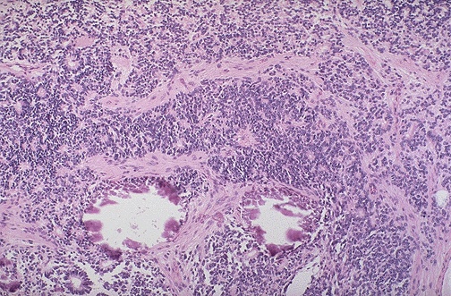
► Retinoblastoma
(2). Retinoblastoma is one of the "small
blue cell tumors" of childhood. Necrosis and dystrophic
calcification are commonly seen within this tumor. At low magnification,
two small calcification can be seen below center.
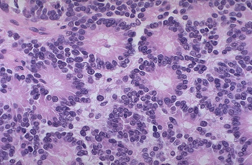
► Retinoblastoma
(3). Retinoblastoma is one of the "small
blue cell tumors" of childhood. Necrosis and dystrophic
calcification are commonly seen within this tumor. The characteristic
microscopic pattern is arrangement of the small blue cells into
Flexner-Wintersteiner "rosettes" as shown here.
|
| |
|
| |
Uniformed
Services U. of the Health Sciences /Bethesda, Maryland/, MedPix™ -
Medical Image Database
|
| |
Eye
and Orbit - Retinoblastoma
|
| |
►
Axial T1W MR demonstrates a mass in the inferior right
retina,
isointense to brain
► Axial
T2W MR demonstrates a mass in the inferior right retina, hypointense to
brain
► Axial
and coronal gadolinium enhanced MR images demonstrates an enhancing mass
in the inferior right retina
► Axial
and coronal gadolinium enhanced MR images demonstrates an enhancing mass
in the inferior right retina
|
| |
Eye
and Orbit - Retinoblastoma
|
| |
► 4
year old girl with "cat's eye". Notice that there is a yellowish
light reflection (leukokoria) from the nasal portion of the patient's
right eye.
► Resected
speciment (enucleation). There is a large intraocular mass, occupying more
than half of the volume of the globe.
► Axial
CT scan. There is a partially calcified intraocular mass in the medial (nasal)
portion of the patient's right eyeball.
► Axial
non-contrast CT scan demonstrates a calcifed mass extending from the left
retina.
► Axial
non-contrast CT scan demonstrates a calcifed mass extending from the left
retina.
|
| |
|
| |
Other
Links:
► World of Ophthalmology - Diseases & Conditions -
Retinoblastoma |
| |
|
|
|
Top of
the Page |
| |
|
| |
Home
Disclaimer
Privacy
Policy
Advertising
Policy
About Me and My Site |
| |
|
| |
'World
of Ophthalmology' has no control over content of these links
If you know of any links appropriate for page, please submit
for inclusion |
|
|
|
|
|
|
|
|
|
|
|
|
|
|
|