World of Ophthalmology Dr. Victor Zamyatin's Personal Web Site
| Ophthalmology - Home Page | Anatomy | Physiology | Diseases | Diagnostic Tests | Surgical Procedures |
| Medications | Contact Lenses & Eyeglasses | Journals | Images | Links | FAQs | News |
| Congresses, Events | Trials | Eye Banks | Equipment | Eye & Vision Companies | Contact |
|
Encyclopaedia of Ophthalmology - Greatest Links' Collection |
|
Look 'World of Ophthalmology' on http://www.wophth.com ! |
|
Advertising
Banners appear in the left column |
|
► Anatomy |
|
► Diseases |
|
► Surgery |
 |
| We subscribe to the HONcode principles . Verify here |
|
'World of Ophthalmology' depends on donations and sponsorship. Sponsors will be offered the opportunity to
advertize/display their brand |
||||||||||||||
| Catalogue of Ophthalmic Images | ||||||||||||||
|
|
||||||||||||||
| Ocular Globe | ||||||||||||||
|
2 Eyes - Anatomy / ► Ocular Globe |
||||||||||||||
| Anatomy Atlases | ||||||||||||||
|
Atlas of Human Anatomy / Plate 31: Visual and hearing apparatus |
||||||||||||||
| ► The
left eye of the dissected face, with its muscles and nerves.
► The tear and eyelid glands of the left eye. ► Both orbital cavities, opened from above, with their muscles and nerves. ► The right ocular bulb in the orbit, cut through the middle, opened from the lateral side. |
||||||||||||||
| ► Section 1. Head and Neck | ||||||||||||||
| FotoWeb - Ophthalmic Images | ||||||||||||||
|
Animations (Movies) |
||||||||||||||
| ► Ocular globe. Basic anatomy | ||||||||||||||
| ► Ocular
arterial vascularisation. Carotid artery, ophthalmic artery, ciliary
arteries, retina central artery.
► Ocular globe layers, muscular insertion of oblique and upper and lower straigths. ► Extrinsic eye muscles. Sagital cut of the orbit. ► Sagital cut of the ocular globe in the orbit. Orbitary fat of the muscular cone. Vitreous cavity. Arrival of the optic nerve to the ocular globe. ► Venous circulation of the ocular globe. ► Eyeball muscles. Inferior view ► Eyeball muscles. Anterior view ► Eyeball muscles. Superior view ► Sagittal cut of the eyeball ► Transverse cut of the head ► Sagittal cut of the eyeball. General view ► Sagittal cut of the eyeball. Detail of the uveal tract ► Sagittal cut of the eyeball. Detail of sclera and optic nerve ► Front cut of the eyeball. Posterior view of the lens.(II) ► Eyeball, posterior view. |
||||||||||||||
| U. of Delaware, Department of Biological Sciences, Mammalian Histology (B408) | ||||||||||||||
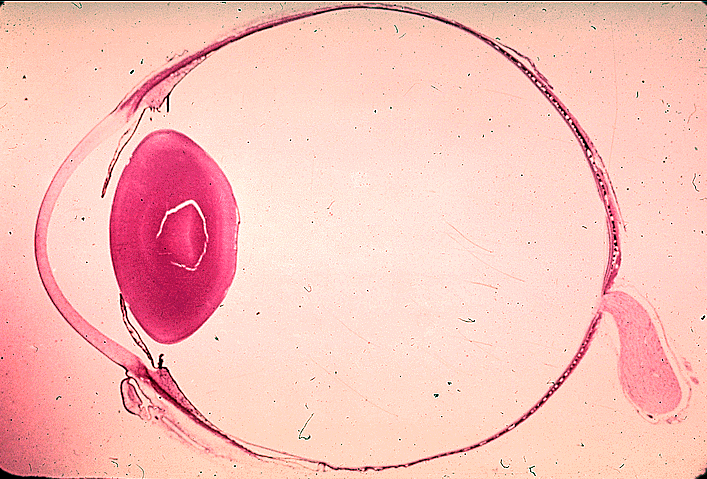
| ► Eyeball Saggital Section, Panoramic | ||||||||||||||
| Other
Links: ► World of Ophthalmology - Eye Anatomy |
||||||||||||||
|
Home Disclaimer Privacy Policy Advertising Policy About Me and My Site |
||||||||||||||
|
'World
of Ophthalmology' has no control over content of these links |
||||||||||||||
Web
site content and design by Dr. Victor Zamyatin
© 2006, World
of Ophthalmology
Contact adress: v.zamyatin@gmail.com