World of Ophthalmology Dr. Victor Zamyatin's Personal Web Site
| Ophthalmology - Home Page | Anatomy | Physiology | Diseases | Diagnostic Tests | Surgical Procedures |
| Medications | Contact Lenses & Eyeglasses | Journals | Images | Links | FAQs | News |
| Congresses, Events | Trials | Eye Banks | Equipment | Eye & Vision Companies | Contact |
|
Encyclopaedia of Ophthalmology - Greatest Links' Collection |
|
Look 'World of Ophthalmology' on http://www.wophth.com ! |
|
Advertising
Banners appear in the left column |
|
► Anatomy |
|
► Diseases |
|
► Surgery |
 |
| We subscribe to the HONcode principles . Verify here |
|
'World of Ophthalmology' depends on donations and sponsorship. Sponsors will be offered the opportunity to
advertize/display their brand |
||||||||||||||||||||
| Catalogue of Ophthalmic Images | ||||||||||||||||||||
|
|
||||||||||||||||||||
| Iris | ||||||||||||||||||||
|
2 Eyes - Anatomy / 2 Uvea / 2 Iris |
||||||||||||||||||||
|
||||||||||||||||||||
| Anatomy Atlases | ||||||||||||||||||||
|
Atlas of Microscopic Anatomy - A Functional Approach / Section 16. Special Senses |
||||||||||||||||||||
| Bartleby.com - Gray’s Anatomy of the Human Body | ||||||||||||||||||||
| ►
FIG.
872– The choroid
and iris.
► FIG. 873– The arteries of the choroid and iris. The greater part of the sclera has been removed. ► FIG. 876– Vessels of the choroid, ciliary processes, adn iris of a child. |
||||||||||||||||||||
| EyeMDLink.com | ||||||||||||||||||||
| ► Iris | ||||||||||||||||||||
| FotoWeb - Ophthalmic Images | ||||||||||||||||||||
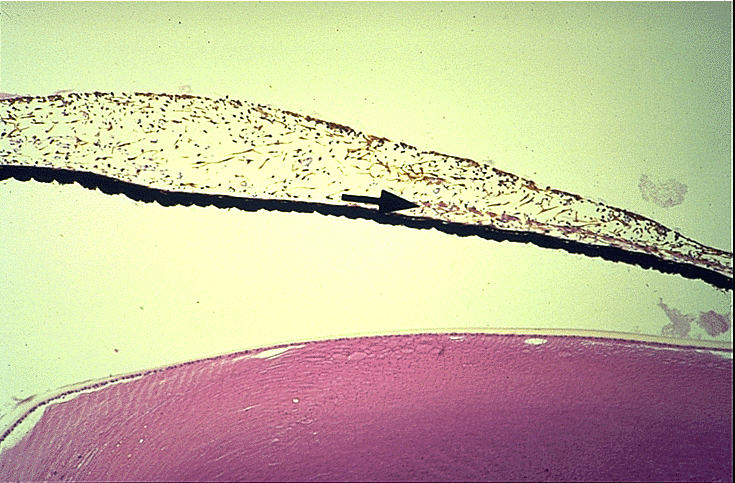
| ►
Uvea,
ciliary body, production and drainage of aqueous body; trabecular
mesh; Schlemm canal, sphincter and radial muscles of the iris.
► Sagital cut of the lower ciliary region showing: the crystalline, zonule fibres, ciliary crown, flat ciliary part, ora serrata, posterior face of the iris, retina... ► Ciliary muscle with its compounds, dissected and exposed: longitudinal, oblique and circulra muscular layers of the cyliary muscle; scleral venous sinus and its collecting veins; pectineal ligament; scleral spur, iris and ciliary crown. |
||||||||||||||||||||
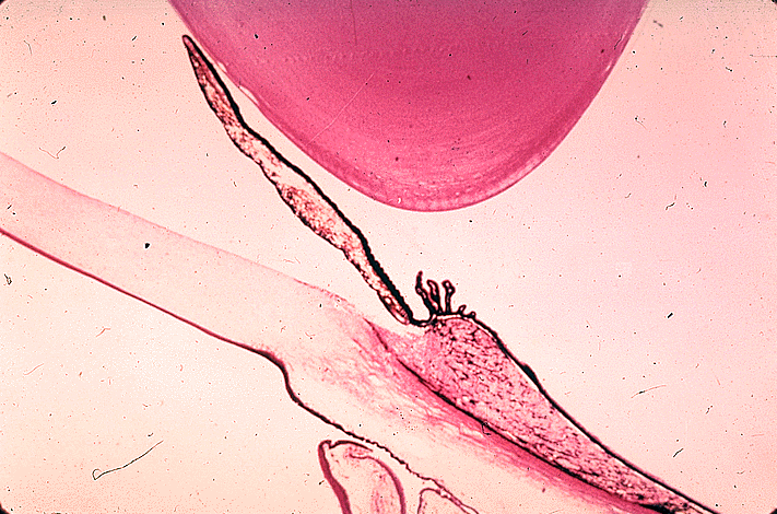
| ► Anterior
and posterior face of the iris.
► Iris section; anterior limiting chamber consisting of intertwisted fibroblasts and posterior one of melanocytes with capillaries running between them and collagen fibres; stroma of laxus bindweb with spiral-type arterioles and venules of ... ► Sagittal cut of the eyeball. Lens |
||||||||||||||||||||
| Northeastern Ohio Universities, College of Medicine, Neurobiology Department | ||||||||||||||||||||
| ► Eye Anatomy / Eye - Retina - Iris | ||||||||||||||||||||
| St Luke's Cataract & Laser Institute - Anatomy Focus | ||||||||||||||||||||
| ► Iris | ||||||||||||||||||||
| Suny Downstate Medical Center | ||||||||||||||||||||
| ► Iris | ||||||||||||||||||||
| U. of Bristol, Bristol Biomedical Image Archive | ||||||||||||||||||||
| ► Fluorescein angiogram of iris | ||||||||||||||||||||
| U. of Delaware, Department of Biological Sciences, Mammalian Histology (B408) | ||||||||||||||||||||
| ► Ciliary Body, Iris, & Lens | ||||||||||||||||||||
| U. of Iowa Health Care - Virtual Hospital - Atlas of Microscopic Anatomy | ||||||||||||||||||||
| ►
Iris and Lens
► Iris |
||||||||||||||||||||
| U. of Kansas Medical Center - Eye & Ear Anatomy and Histology | ||||||||||||||||||||
| ►
8.
Iris
► 9. Iris |
||||||||||||||||||||
| U. of North Carolina /Chapel Hill/, School of Medicine, Embryo Images Online - Eye Development | ||||||||||||||||||||
| ► IV. Iris, Ciliary Body (Weeks 9-15) | ||||||||||||||||||||
| U. of Texas Medical Branch, Cell Biology Graduate Program, Microanatomy Web Atlas | ||||||||||||||||||||
| ► Cornea, Iris and Lens | ||||||||||||||||||||
| Vision CyberMedia Group, Development of the Eye Site | ||||||||||||||||||||
| ► Figure 5.
Successive sagittal sections of developing eye at: A, 5 weeks B, 6 weeks C, 20 weeks
D, Newborn
► Figure 3. Sagittal section through the developing eye at about seven weeks. ► Figure 10. A, B, Development of the rim of the optic cup into the iris and ciliary body. ► Figure 11. Sagittal section through the developing eye at 15 weeks. |
||||||||||||||||||||
| Xalatan Image Library | ||||||||||||||||||||
| ► The Iris | ||||||||||||||||||||
| ►
The Iris
► The Iris ► The Iris Dilator and Sphincter Muscles |
||||||||||||||||||||
| ► The Iris and Ciliary Body - Anatomy | ||||||||||||||||||||
| Other
Links: ► World of Ophthalmology - Eye Anatomy - Iris |
||||||||||||||||||||
|
Home Disclaimer Privacy Policy Advertising Policy About Me and My Site |
||||||||||||||||||||
|
'World
of Ophthalmology' has no control over content of these links |
||||||||||||||||||||
Web
site content and design by Dr. Victor Zamyatin
© 2006, World
of Ophthalmology
Contact adress: v.zamyatin@gmail.com