World of Ophthalmology Dr. Victor Zamyatin's Personal Web Site
| Ophthalmology - Home Page | Anatomy | Physiology | Diseases | Diagnostic Tests | Surgical Procedures |
| Medications | Contact Lenses & Eyeglasses | Journals | Images | Links | FAQs | News |
| Congresses, Events | Trials | Eye Banks | Equipment | Eye & Vision Companies | Contact |
|
Encyclopaedia of Ophthalmology - Greatest Links' Collection |
|
Look 'World of Ophthalmology' on http://www.wophth.com ! |
|
Advertising
Banners appear in the left column |
|
► Anatomy |
|
► Diseases |
|
► Surgery |
 |
| We subscribe to the HONcode principles . Verify here |
|
'World of Ophthalmology' depends on donations and sponsorship. Sponsors will be offered the opportunity to
advertize/display their brand |
||||||||||||||||||||
| Catalogue of Ophthalmic Images | ||||||||||||||||||||
|
|
||||||||||||||||||||
| Macula Lutea | ||||||||||||||||||||
|
2 Eyes - Anatomy / 2 Retina / 2 Macula Lutea |
||||||||||||||||||||
|
||||||||||||||||||||
| American Health Assistance Foundation | ||||||||||||||||||||
| ► Normal Macula | ||||||||||||||||||||
| EyeMDLink.com | ||||||||||||||||||||
| ► Macula | ||||||||||||||||||||
| FotoWeb - Ophthalmic Images | ||||||||||||||||||||
| ►
Normal
eye bottom. Normal papilla with ratio P/E = 0.3.
► Arteries and veins of the retina in a normal eye bottom. Viewing of the macula and description of the main vascular trunks and their nasal and temporal branches. ► Normal young eye bottom. Typical for children and teenagers. Reflections produced by the internal limiting layer of the retina and the tortuosity of the arteries and veins may be observed. ► Normal eye bottom of young adult. Reflections disappear and the fine velvet of the retina may be observed. The vessels may still be seen as tortuous. ► Normal eye bottom of a 75 years old person. The retina is thinned, vessels are rectilinear and a tabby choroid may be observed. ► Eye fundus in miopia ► Normal eye bottom |
||||||||||||||||||||
| Northeastern Ohio Universities, College of Medicine, Neurobiology Department | ||||||||||||||||||||
| ► Eye
Anatomy / Eye - Retina - Macula Lutea
► Eye Anatomy / Eye - Retina - Macula Lutea (Fundoscopic View) ► Eye Anatomy / Eye - Retina - Rod and Cones & Pigmented Epithelium |
||||||||||||||||||||
| Retina and Vitreous of Texas Web Site | ||||||||||||||||||||
| ► A Brief Eye Anatomy Class | ||||||||||||||||||||
| Rochester Institute of Technology, Center for Imaging Science, Dr. Ethan D. Montag Web Site | ||||||||||||||||||||
| ► Parts of the Eye / Retina | ||||||||||||||||||||
| St Luke's Cataract & Laser Institute - Anatomy Focus | ||||||||||||||||||||
| ► Macula | ||||||||||||||||||||
| Vitreo-Retinal Consultants | ||||||||||||||||||||
| ►
The Macula and the Fovea |
||||||||||||||||||||
| Washington Academy of Eye Physicians and Surgeons | ||||||||||||||||||||
| ► Macula / Fovea | ||||||||||||||||||||
| WebVision - The Organization of the Vertebrate Retina (U. of Utah /US/) | ||||||||||||||||||||
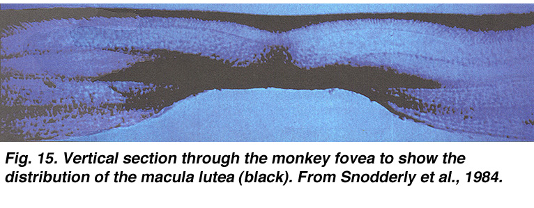
| ►
Ophthalmoscopic
appearance of the retina to show macula lutea (39 K jpeg image)
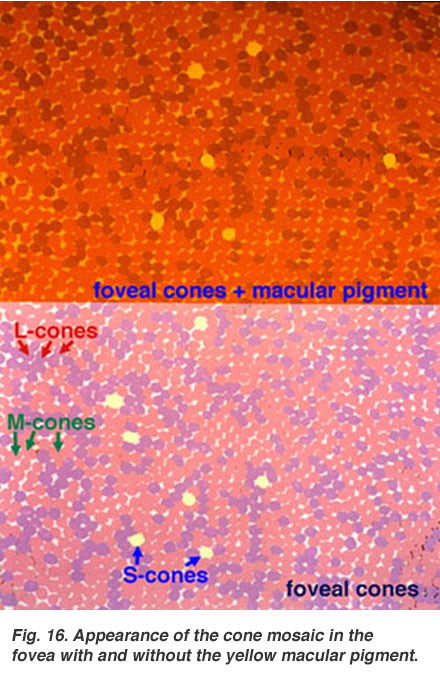
► Appearance of the cone mosaic in the fovea with and without macula lutea (59 K jpeg image) |
||||||||||||||||||||
| Xalatan Image Library | ||||||||||||||||||||
| ► The Macula | ||||||||||||||||||||
| ► Macula Degeneration - Macula | ||||||||||||||||||||
| Other
Links: ► World of Ophthalmology - Eye Anatomy - Macula Lutea |
||||||||||||||||||||
|
Home Disclaimer Privacy Policy Advertising Policy About Me and My Site |
||||||||||||||||||||
|
'World
of Ophthalmology' has no control over content of these links |
||||||||||||||||||||
Web
site content and design by Dr. Victor Zamyatin
© 2006, World
of Ophthalmology
Contact adress: v.zamyatin@gmail.com