|
'World
of Ophthalmology' depends on donations and sponsorship.
Sponsors will be offered the opportunity to
advertize/display their brand
throughout the site, for example |Company
Name|
World of Ophthalmology
|
| |
|
|
Catalogue
of Ophthalmic Images |
| |
|
| |
|
| |
|
|
Anatomy |
|
2 Eyes
- Anatomy
/
2
Anatomy
|
| |
|
|
|
|
|
|
<
Prev |
|
1
2
3 |
|
|
| |
|
| |
National
Eye Institute /US/: Photograph and Image Catalog
|
| |
Normal Eye Anatomy
|
| |
► Eye diagram with
labels.
► Normal
fundus.
► Eye
diagram.
► Anatomy of the
eye.
► Before and after the pupil is
dilated.
► An artist's conception of the interior of a normally
developing premature infant eye.
|
| |
|
| |
Pendleton
Eye Clinic
|
| |
► From Light to
Vision: Anatomy of the
Eye
|
| |
|
| |
Rochester Institute of
Technology, Center for Imaging Science, Dr. Ethan D.
Montag Web Site
|
| |
► Parts of
the Eye
|
| |
|
| |
Suny Downstate Medical Center
|
| |
► Orbits
and Eye
|
| |
|
| |
Syndicat
National des Ophtalmologistes
de France
|
| |
► Anatomie
de l'oeil normal
► Embryologie
de l'oeil
|
| |
|
| |
U. of Kansas Medical
Center - Eye
& Ear Anatomy and Histology
|
| |
► 1.
Eye diagram
|
| |
|
| |
U. of North Carolina
/Chapel Hill/, School of Medicine, Embryo Images Online - Eye Development
|
| |
► I. Eye Fields-Optic Vesicle
(Weeks
3-4)
► II. Optic
Cup, Lens Vesicle,
Choroid Fissure, Hyaloid
Artery (Weeks
5-6)
► III.
Cornea, Anterior Chamber,
Pupillary Membrane, Lens,
Retina (Weeks
7-8)
► IV.
Iris, Ciliary Body (Weeks
9-15)
► V. Eyelids
(Weeks
8-10)
|
| |
|
| |
Vision Correction Website
|
| |
► Anatomy of an Eye
|
| |
|
| |
Vision CyberMedia
Group, Development of the Eye Site
|
| |
Overview
|
| |
► Figure 1.
A, Dorsal cranial view of embryo at about 22
days. B,
Transverse section of an optic sulcus
(groove).
► Figure 2.
A,B,C, Successive schematic sections of developing optic cup and lens
vesicle. D,
Transverse section of optic stalk
► Figure 3.
Sagittal section through the developing eye at about seven weeks.
►►►
More
Images
|
| |
|
| |
Washington
U. in St. Louis, School of Medicine, Neuroscience
Tutorial
|
| |
► Eye
and retina
|
| |
|
| |
WebVision - The
Organization of the Vertebrate Retina (U. of Utah /US/)
|
| |
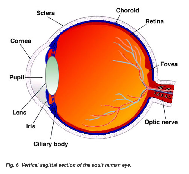
Gross Anatomy of the
Eye
|
| |
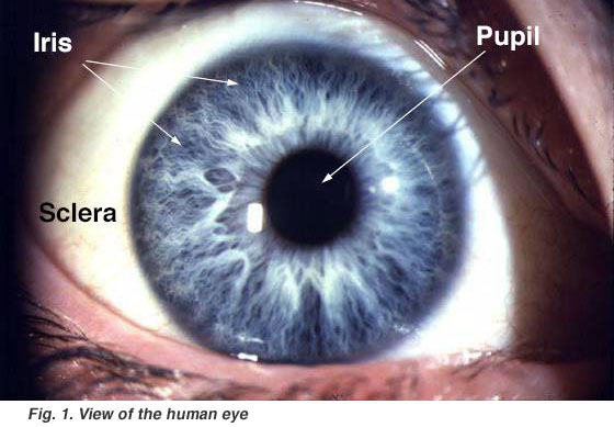
► View
of the human eye (59 K jpeg image)
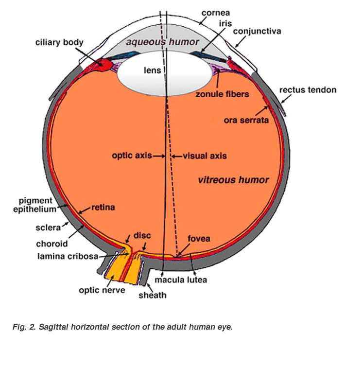
► Sagittal
section of the adult human eye(59 K jpeg image)
► Development
of the eye (59 K jpeg image)
► Morph
of development (683 K quicktime movie)
► Sagittal
section of the adult human eye (59 K jpeg image)
|
| |
|
| |
Xalatan Image
Library
|
| |
Anatomy & Physiology - page 9
|
| |
► The Protective and Supporting
Apparatus of the eye
► The Protective and Supporting
Apparatus of the eye
► The Protective and Supporting
Apparatus of the eye
►►►
More
Images
|
| |
|
| |
York
U., The
Joy of Visual Perception: A Web Book
|
| |
► Master
Diagram of the Eye
► Eye
Cross Section
|
| |
|
| |
Other
Links:
► World of Ophthalmology - Eye Anatomy |
| |
|
|
|
|
|
|
<
Prev |
|
1
2
3 |
|
Top of
the Page |
| |
|
| |
Home
Disclaimer
Privacy
Policy
Advertising
Policy
About Me and My Site |
| |
|
| |
'World
of Ophthalmology' has no control over content of these links
If you know of any links appropriate for page, please submit
for inclusion |
|
|
|
|
|
|
|
|
|
|
|
|
|
|
|